Welcome to Our Biochemistry Lab!
Our biochemistry lab offers a dynamic environment for learning and discovery, equipped with cutting-edge instruments to support advanced research and hands-on training. We provide:
Join us to explore the exciting world of biochemistry with the support of exceptional resources and a collaborative learning atmosphere!
Selected Instruments are shown below-

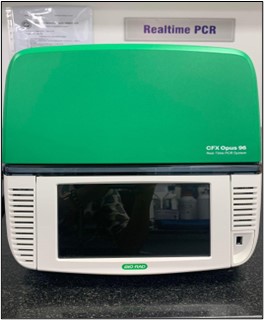
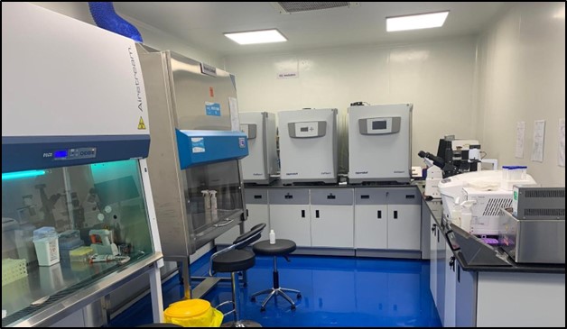
A mammalian cell culture lab is equipped to support the growth and maintenance of mammalian cells in vitro under sterile and controlled conditions. Essential tools include CO₂ incubators for temperature and gas regulation, laminar flow hoods for aseptic handling, and cryostorage for preserving cell lines. Routine activities involve passaging cells, monitoring cell health, and conducting experiments such as drug testing or gene expression studies. Advanced equipment like centrifuges and microscopes supports specialized workflows. The lab is designed to minimize contamination risks while ensuring optimal cell viability, with dedicated areas for media preparation, waste disposal, and equipment storage. Some of the state of the art microscopes in the lab are:
The Zeiss fluorescence microscope, paired with the Axiocam 202 camera, enables high-resolution imaging of fluorescently labeled cellular structures. The camera’s sensitivity ensures detailed visualization and documentation. This setup is ideal for studying protein localization, cellular dynamics, and intracellular processes, offering a powerful tool for advanced cellular and molecular biology research.
An inverted microscope, with objectives placed below the stage, is essential for observing cells in culture flasks or plates without disrupting their environment. It supports routine tasks like confluency checks and cell morphology monitoring. Its compatibility with live-cell imaging makes it indispensable for mammalian cell culture workflows.
The Leica Stellaris 5 DMiB confocal microscope features a spectral detector for precise multicolor imaging and a White Light Laser (WLL) for continuous tunability, allowing flexibility in fluorophore excitation. It supports live-cell imaging with temperature and CO2 control, and its adaptive optics improve image clarity by compensating for tissue aberrations. The system is optimized for high-throughput imaging, offering fast acquisition and multi-position scanning capabilities.
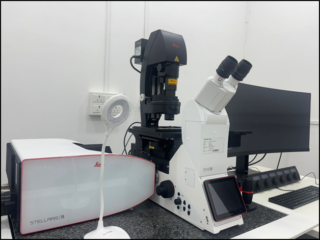

SRM Division of Medical Research, Medical & Health Sciences, SRMIST.
Phone: +914447432591
Email: medresearch@srmist.edu.in
Copyright @ 2026. All Rights Reserved.
